MIT Researchers Build Striatum Cell Atlas to Guide Brain-Disorder Research
An MIT-led study used single-cell RNA sequencing and spatial mapping to identify 31 neuronal subgroups in the human striatum, a brain region involved in movement, decision-making, habits, and reward. The atlas highlighted cell populations linked to addiction, depression, and schizophrenia, while also revealing differences between human and mouse neurons relevant to Huntington’s disease and opioid research.
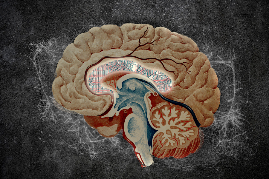
MIT researchers have created a detailed atlas of neurons in the striatum, a brain region involved in movement, decision-making, habit formation, and reward processing. The study, published in Cell, identified 31 neuronal subgroups and pointed to cell populations that may help explain vulnerabilities in Huntington’s disease and differences in drug responses linked to schizophrenia, depression, and substance use disorders.
The striatum is located deep within the brain and receives signals from several regions, including the cortex, midbrain, and hippocampus. The researchers focused on medium spiny neurons, the most common cell type in the striatum. These inhibitory neurons respond to dopamine and are broadly divided into direct and indirect pathways, associated with promoting or suppressing movement. They are also found in more specialized populations, particularly in the lower, or ventral, striatum.
To examine that cellular diversity, the team analyzed postmortem human striatal tissue representing different anatomical regions. Brain banks in the United States and Canada contributed samples. The researchers combined single-cell RNA sequencing with multiplexed fluorescent in situ hybridization and spatial transcriptomics. Together, the methods showed both which genes individual cells expressed and how different cell populations were arranged within the tissue.
The analysis identified nine types of medium spiny neurons among the 31 neuronal groups. Two populations stood out because of their apparent relevance to psychiatric and substance-use research. The D1 outlier population expressed high levels of genes associated with addiction and substance use disorder, including genes related to opioid response. The D2 outlier population expressed genes that respond to antidepressants. Both groups appeared to respond strongly to clozapine, an antipsychotic medication used in schizophrenia treatment.
The findings may help researchers investigate whether drugs can be directed more selectively toward particular neuronal populations. MIT’s Myriam Heiman said the results could provide a basis for studying treatments that address psychosis while avoiding clozapine’s serious blood-related risk. The study did not establish a new treatment or show that targeting these cells is safe or effective in patients.
The atlas also offered a possible explanation for why the upper, or dorsal, striatum is especially vulnerable to Huntington’s disease. Dorsal medium spiny neurons expressed higher levels of MSH2 and MSH3, genes involved in increasing the number of CAG repeats in the huntingtin gene. The researchers reported that accumulation of these repeats can make the mutated huntingtin protein more harmful to cells.
By contrast, a rare group of medium spiny neurons forming island-like structures in the ventral striatum appeared more resistant to CAG-repeat accumulation. The researchers said studying the genes expressed—or not expressed—by these cells could provide clues about how to make other neurons more resilient. That possibility remains a research direction rather than a demonstrated therapy.
The team also compared human tissue with mouse samples and found differences in genes related to drug response and substance use disorders. The gene encoding the mu opioid receptor, OPRM1, was highly expressed in the human D1 outlier population but not in the corresponding mouse population. The result suggests that some standard mouse models may not fully reproduce human opioid-response biology, according to the researchers.
The study’s atlas was assembled from donated human brain tissue and collaboration across institutions. Its main contribution is a map of cellular diversity and regional vulnerability in the striatum, which researchers can use to frame future studies of Huntington’s disease, opioid use disorder, schizophrenia, depression, and addiction.
Reporting Note
Biohack Report distinguishes preliminary findings, clinical evidence and commercial claims whenever the available reporting supports that distinction. Coverage is informational and is not medical advice.