UCLA Studies Identify Metabolic and Physical Signals Guiding Brain Development
Two UCLA studies report that radial glia, stem cells involved in building the human cerebral cortex, respond to both glucose-processing pathways and physical contact from thalamic projections. The findings, based on donated tissue, brain organoids and stem-cell-derived assembloids, offer new clues about how early developmental signals influence neuron production.
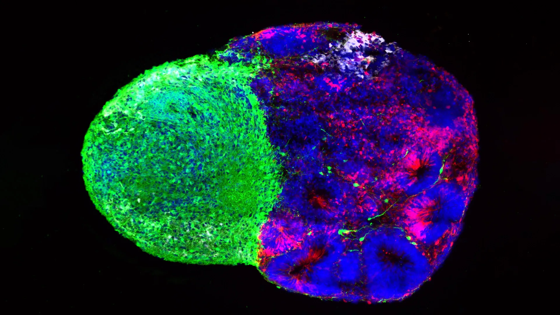
Two UCLA studies suggest that early human brain development is guided by more than genetic instructions alone. Radial glia—the stem cells that generate many neurons and support cells in the cerebral cortex—also respond to metabolic conditions and physical signals from other parts of the developing brain.
In research published in Cell, investigators created a metabolic map of the developing human cortex using donated human tissue and brain organoids grown from stem cells. The work identified the pentose phosphate pathway, a glucose-processing route involved in supplying materials for rapidly dividing cells, as an important part of radial glia behavior.
When researchers reduced glucose availability or disrupted that pathway, the radial glia changed the types of cells they produced. The models generated more inhibitory neurons and other cell types that typically emerge later in development. The result indicates that metabolism may actively influence cell-fate decisions rather than simply provide background energy for growth.
The researchers said the findings could help investigations into how maternal nutrition, metabolic disorders and other environmental factors affect the developing brain. The metabolic atlas was also described as a resource for studying metabolism during human brain formation.
A separate study, published in Science and led by Claudia Nguyen, examined signals from the thalamus, a brain structure involved in relaying information through the nervous system. In human stem-cell-derived brain assembloids—models made by combining developing brain tissues—researchers found that thalamic projections physically contacted radial glia before the final connections between thalamic and cortical neurons had formed.
That contact altered the stem cells’ behavior, leading them to produce more excitatory neurons, including upper-layer neurons that are particularly expanded in the human brain. The researchers said this direct point of contact had not previously been identified and may not occur in rodents, according to the source material.
The study also examined NRXN1, a gene involved in neuronal connections and previously associated with autism spectrum disorder. Assembloids made from patient-derived cells carrying an NRXN1 mutation showed altered thalamic signaling. Those changes affected the balance between the number of radial glia and the neurons they generated.
Together, the studies portray radial glia as highly responsive to their surroundings. One set of experiments focused on nutrient processing, while the other investigated physical cellular contact. Both relied in part on laboratory models, including organoids and assembloids, so they provide insight into developmental mechanisms rather than evidence that changing these signals can treat a condition in people. The researchers said the findings may help clarify normal brain development, vulnerability to neurodevelopmental disorders and related cellular programs in brain cancer.
Reporting Note
Biohack Report distinguishes preliminary findings, clinical evidence and commercial claims whenever the available reporting supports that distinction. Coverage is informational and is not medical advice.